
The Protein Data Bank (PDB) is a repository for the 3-D structural data of large biological molecules, such as proteins and nucleic acids. (See also crystallographic database). The data, typically obtained by X-ray crystallography or NMR spectroscopybiologists and biochemists from around the world, are freely accessible on the Internet via the websites of its member organisations (PDBe, PDBj, and RCSB). The PDB is overseen by an organization called the Worldwide Protein Data Bank,
The PDB is a key resource in areas of structural biology, such as structural genomics. Most major scientific journals, and some funding agencies, such as the NIH in the USA, now require scientists to submit their structure data to the PDB. If the contents of the PDB are thought of as primary data, then there are hundreds of derived (i.e., secondary) databases that categorize the data differently. For example, both SCOP and CATH categorize structures according to type of structure and assumed evolutionary relations; GO categorize structures based on genes.
The PDB originated as a grassroots effort.In 1971, Walter Hamilton of the Brookhaven National Laboratory agreed to set up the data bank at Brookhaven. Upon Hamilton's death in 1973, Tom Koeztle took over direction of the PDB. In January 1994, Joel Sussman was appointed head of the PDB. In October 1998,[2] the PDB was transferred to the Research Collaboratory for Structural Bioinformatics (RCSB); the transfer was completed in June 1999. The new director was Helen M. Berman of Rutgers University (one of the member institutions of the RCSB).[3] In 2003, with the formation of the wwPDB, the PDB became an international organization. The founding members are PDBe (Europe), RCSB(USA), and PDBj (Japan). The BMRB joined in 2006. Each of the four members of wwPDB can act as deposition, data processing and distribution centers for PDB data. The data processing refers to the fact that wwPDB staff review and annotates each submitted entry. The data are then automatically checked for plausibility. (The source code for this validation software has been made available to the public at no charge.
The file format initially used by the PDB was called the PDB file format. This original format was restricted by the width of computer punch cards to 80 characters per line. Around 1996, the "macromolecular Crystallographic Information file" format, mmCIF, started to be phased in. An XML version of this format, called PDBML, was described in 2005.The structure files can be downloaded in any of these three formats. In fact, individual files are easily downloaded into graphics packages using web addresses:
- For PDB format files, use, e.g.,
http://www.pdb.org/pdb/files/4hhb.pdb.gz or http://pdbe.org/download/4hhb - For PDBML (XML) files, use, e.g.,
http://www.pdb.org/pdb/files/4hhb.xml.gz or http://pdbe.org/pdbml/4hhb
The "
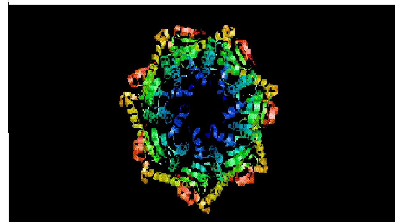
4hhb" is the PDB identifier. Each structure published in PDB receives a four-character alphanumeric identifier, its PDB ID. (This cannot be used as an identifier for biomolecules, because often several structures for the same molecule—in different environments or conformations—are contained in PDB with different PDB IDs.)HtrA
H198P/T167V DOUBLE MUTANT OF DEGS-DELTAPDZ PROTEASE
H198P/T167V DOUBLE MUTANT OF DEGS-DELTAPDZ PROTEASE
Primary Citation |

LonA
CRYSTAL STRUCTURE OF BACILLUS SUBTILIS LON N-TEMINAL DOMAIN
CRYSTAL STRUCTURE OF BACILLUS SUBTILIS LON N-TEMINAL DOMAIN
Primary Citation

Primary Citation

0 comments:
Post a Comment